Specialty MRI Services
From "Head to Toe"
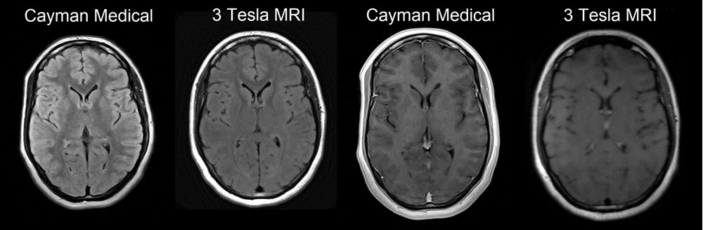
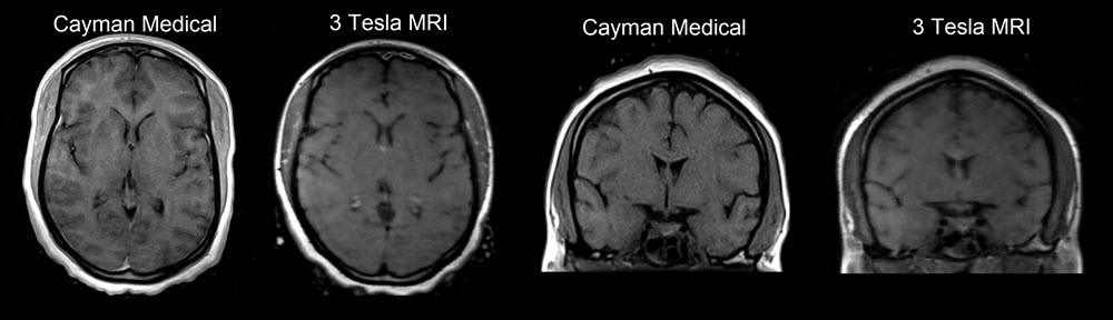
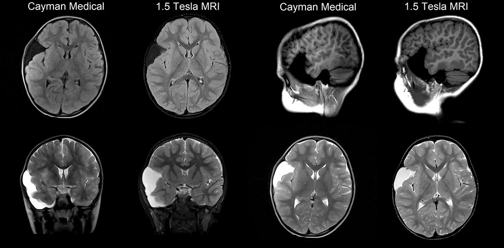
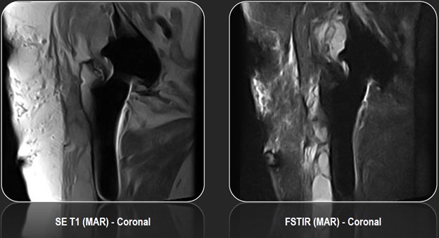
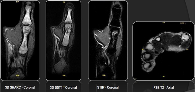
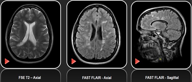
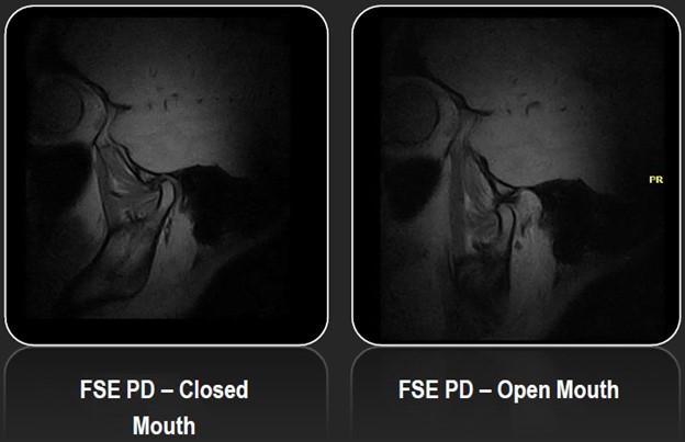
Head & Neck Imaging
Brain, Orbits, Internal Aditory Canal (IAC), Sella (Pituitary), Temporomandibular Joints (TMJ), Neck
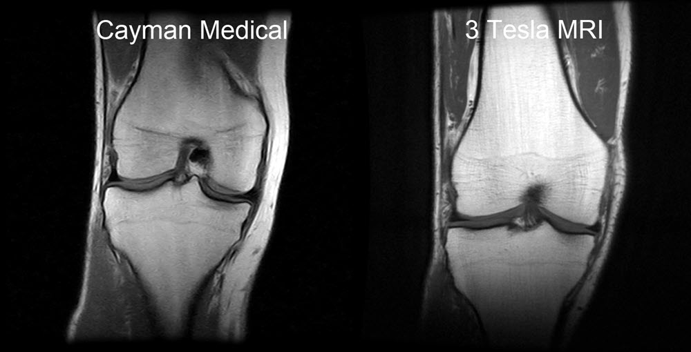
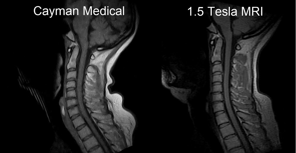
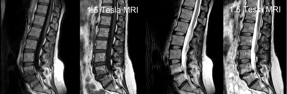
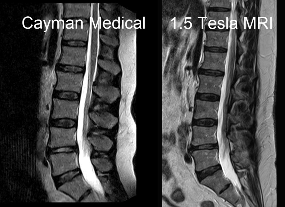
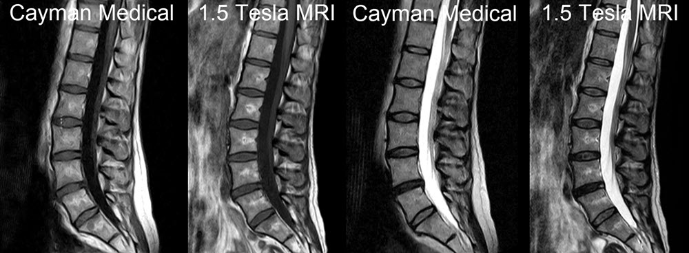
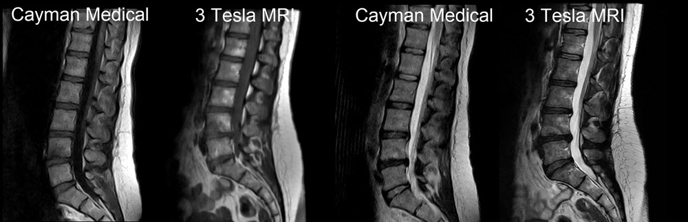
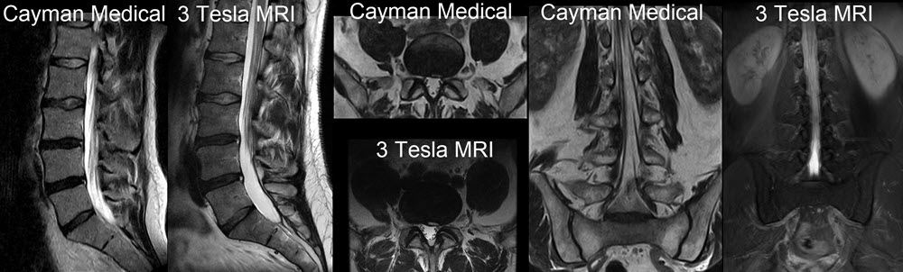
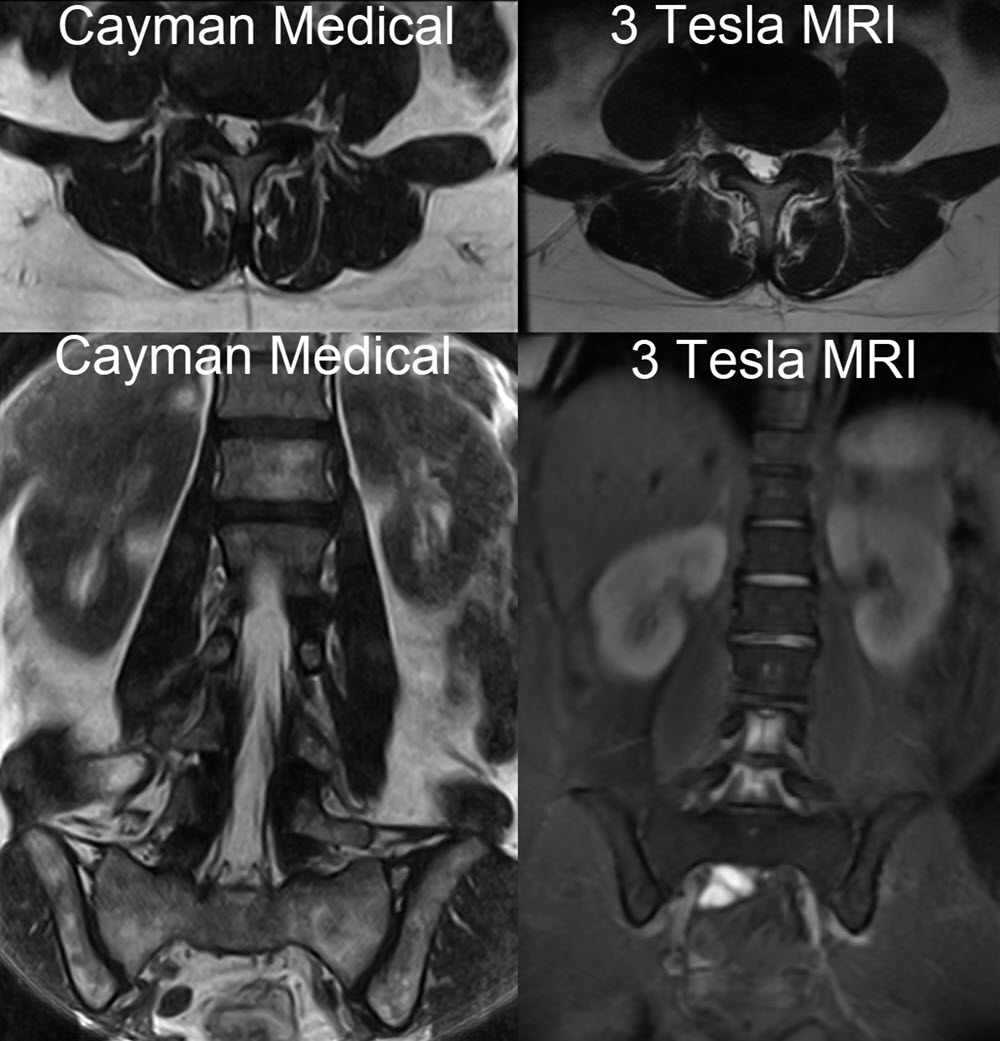
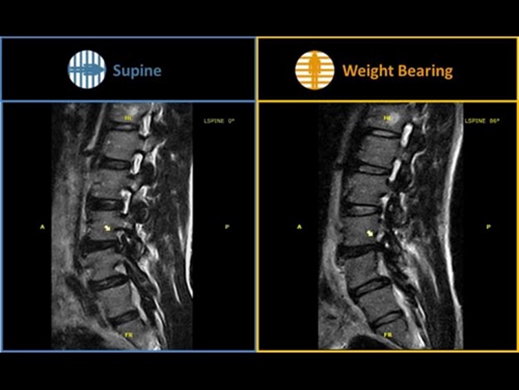
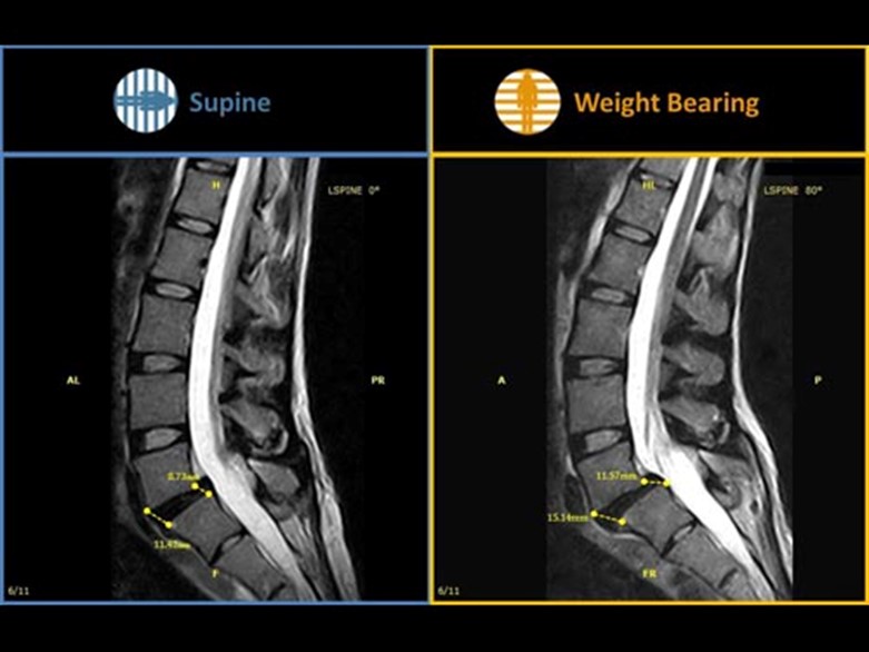
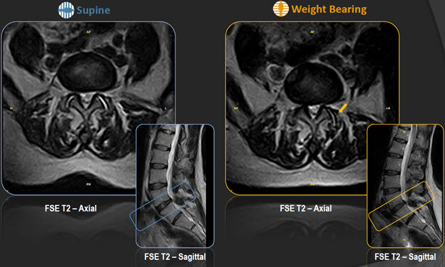
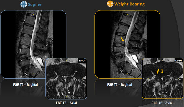
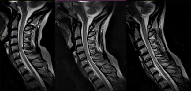
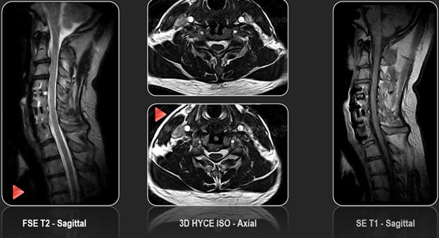
Spine Imaging
Craniocervical Junction, Cervical Spine, Thoracic Spine, Lumbar Spine, Sacrum, Sacroiliac joints